The Power of Photogrammetry in Full-Arch Implant Restorations
Advances in digital technology have transformed dentistry, bringing greater precision, reduced chair time, and enhanced patient comfort. Among the innovations driving this progress, photogrammetry has become a game-changer for full-arch implant restorations, one of the most complex areas of implant dentistry.
The Rise of Digital Dentistry
In implant dentistry, digital workflows have introduced techniques that elevate the accuracy and efficiency of prosthetic results. Full-arch restorations, known for their complexity, benefit immensely from cutting-edge solutions like photogrammetry and 3D printing. These technologies streamline procedures, improve outcomes, and set new standards for precision.
Why Precision Matters in Full-Arch Restorations
Achieving a passive fit is essential in full-arch implant restorations. Each stage of the prosthetic process can introduce small errors, which, if left unchecked, may lead to a poor fit. This can cause strain on implants and connectors, potentially leading to complications. The goal is to reduce marginal gaps as much as possible, ensuring a passive fit that minimizes stress and increases the likelihood of long-term success.
How Photogrammetry Works
Photogrammetry is a transformative technology that captures highly accurate, three-dimensional representations of dental implant positions. It uses a series of two-dimensional images to digitally map the precise positioning and angulation of implants.
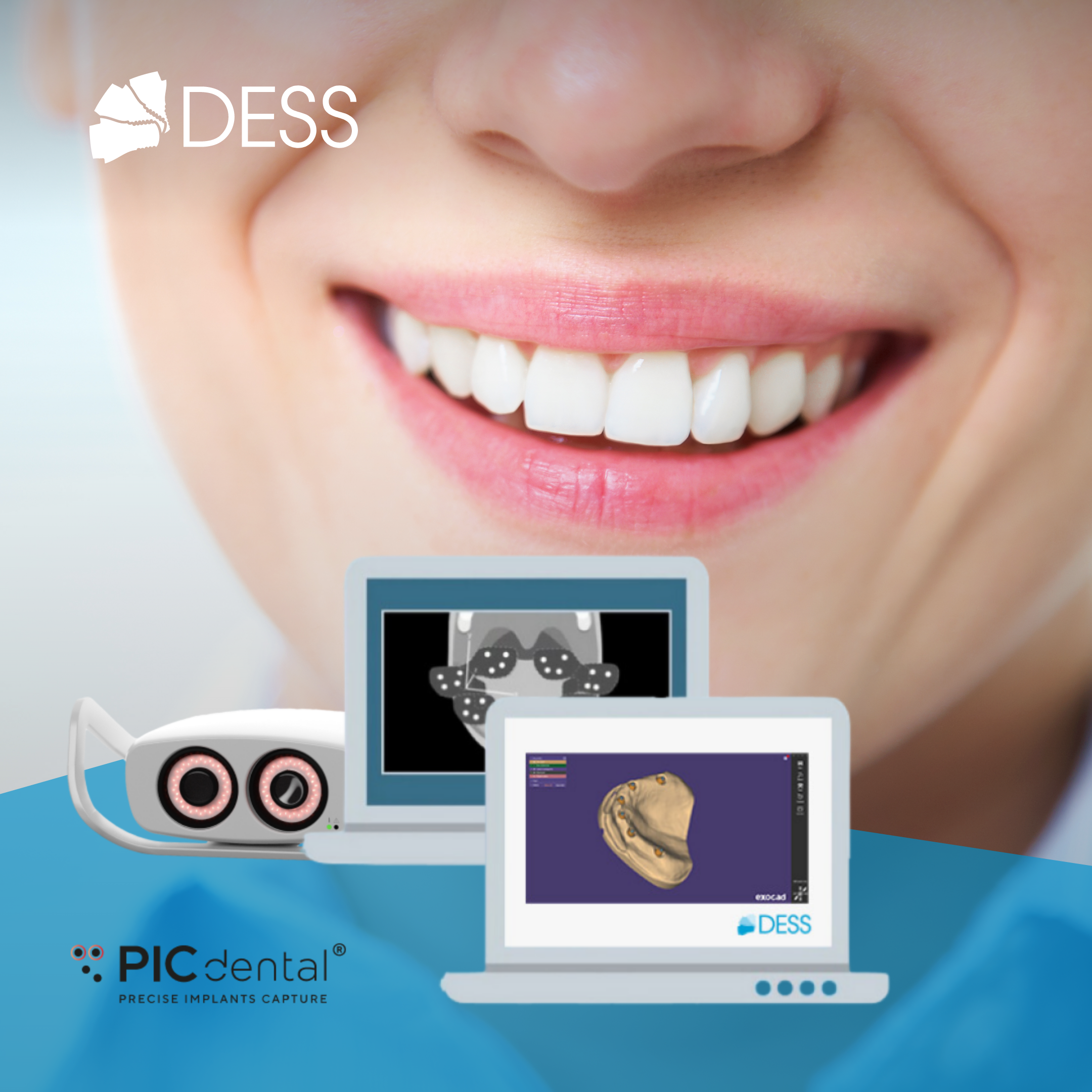
In implant dentistry, photogrammetry involves connecting photogrammetry-coded abutments to the implants. A specialized camera, like the PIC camera®, captures multiple images of these abutments from different angles. The system’s software then processes these images, using mathematical algorithms to create a 3D digital representation of the implant positions. This non-contact method ensures unmatched accuracy without the invasiveness of traditional impression techniques.
Photogrammetry vs. Conventional Methods
Photogrammetry offers significant advantages over traditional methods like intraoral scanning or physical impressions. It is faster, more accurate, and less invasive. Studies show that photogrammetry delivers superior results in capturing implant positions, making it ideal for complex cases like full-arch restorations.
The world’s first commercial dental photogrammetry solution, the PIC system, set the standard for accuracy, and it has the most peer-reviewed scientific evidence validating its performance. Today, other systems like iMetric iCam4D and MicronMapper also provide advanced solutions in implant dentistry.
Integrating Photogrammetry with Intraoral Scanning
While photogrammetry captures precise implant positions, intraoral scanning still plays a vital role in recording soft tissue details like the gingiva. Combining these two technologies results in a digital master model that includes both implant and soft tissue geometry. This integration leads to a highly accurate, predictable fit in the final prosthesis.
DESS® and PIC dental® have partnered to offer a comprehensive digital workflow, utilizing the PIC system for precise implant mapping and DESS® abutments for soft tissue scanning and CAD design. This combination ensures a passive fit, enhancing the success of full-arch restorations.
Precision and Workflow Efficiency with the PIC System
The PIC system, known for its accuracy, uses a stereo camera to capture detailed images that translate into precise implant positions. Under controlled conditions, it’s accuracy is down to 4 microns, making it the most precise dental photogrammetry system available. With over 1 million successful clinical cases and numerous studies backing its efficacy, the PIC system remains the industry leader.
Streamlined Workflow: DESS® and PIC dental®
The collaboration between DESS® and PIC dental® creates a seamless digital workflow for full-arch restorations:
1. Photogrammetry-Coded Abutments: PIC transfers are connected to the patient’s implants. | |
2. Image Capture: The PIC camera® captures images of the abutments. | |
3. Data Processing: PIC suite software calculates exact implant positions and exports the data as an STL file. | |
4. Soft Tissue Scanning: DESS® abutments are used for intraoral scanning, capturing soft tissue details. | |
5. File Integration: Both the implant and soft tissue data are combined in CAD software like Exocad or 3Shape to design the final prosthesis. |
Enhancing Outcomes with DESS® Solutions
DESS® offers a wide range of options to further improve your full-arch restorations. Among these is the new Full Arch Screw for Multi-Unit abutments, featuring a thicker structure seat thickness and the ability to correct the screw channel up to 25 degrees. This innovative solution is integrated into DESS® Libraries.
The combination of mandibular and soft tissue impressions taken with the intraoral scanner, along with the precise 3D representations of implant positions from photogrammetry, enables the creation of highly accurate CAD designs.
DESS® Libraries, compatible with PIC dental®, support seamless integration with both DESS® Abutments and PIC files in CAD software like Exocad and 3Shape. You can download them here.
Elevate Your Full-Arch Practice with DESS® and PIC dental®
By integrating the precision of the PIC system and DESS® abutments, you can achieve unparalleled accuracy in full-arch implant restorations. This collaboration revolutionizes your workflow, improves patient satisfaction, and enhances clinical efficiency. Ready to elevate your practice? Explore the benefits of photogrammetry with DESS® and PIC dental® today.
(1)https://www.sciencedirect.com/topics/agricultural-and-biological-sciences/photogrammetry


